
Procedimientos
En Southeastern Neurosurgical Specialists, nos enorgullece ofrecer una gama completa de servicios a nuestros pacientes para el tratamiento de diversas afecciones. Nuestros profesionales altamente capacitados cuentan con años de experiencia en el diagnóstico y tratamiento de estas afecciones, y estaremos encantados de ayudarle a encontrar la solución que mejor se adapte a sus necesidades individuales.
Nos esforzamos por crear planes de tratamiento individualizados para cada uno de nuestros pacientes, ya que cada persona tiene una historia y necesidades únicas. Algunos de nuestros servicios incluyen:
Procedimientos craneales
Procedimientos cervicales
Procedimientos lumbares
Deformidad espinal / Escoliosis en adultos
Nervio periférico
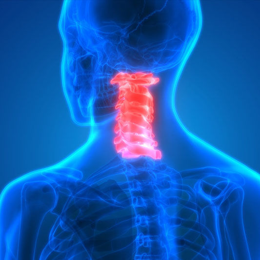
Procedimientos cervicales
Discectomía cervical anterior con fusión (ACDF): procedimiento de fusión en el que se accede a la columna vertebral a través de la parte frontal del cuello. La fusión cervical anterior es una operación que se realiza en la columna superior para aliviar la presión sobre una o más raíces nerviosas o sobre la médula espinal. El término deriva de las palabras anterior (frontal), cervical (cuello) y fusión (unión de las vértebras mediante un injerto óseo).
¿Qué es una discectomía cervical anterior con fusión (ACDF)?
Su profesional de la salud podría recomendarle una ACDF si padece una hernia discal o degeneración en la parte superior de la columna vertebral, conocida como zona cervical.La discectomía cervical anterior es un procedimiento quirúrgico que consiste en acceder a la parte frontal (anterior) del cuello (cervical) y extirpar un disco cervical dañado (discectomía). Se colocan implantes de hueso y/o metal en lugar del disco dañado para fusionar las dos vértebras.
¿Quién necesita este procedimiento?
Si usted presenta alguna de las siguientes afecciones o síntomas, podría ser candidato para una ACDF:Debilidad en la mano o el brazo.
Dolor en el brazo más intenso que el dolor de cuello.
Entumecimiento/debilidad en brazos y extremidades
Discos degenerativos o hernias discales
Otros síntomas cervicales que no han respondido a la medicación o a la fisioterapia.
Su médico podrá revisar los síntomas y las causas exactas que le afectan y por qué usted podría ser candidato para la discectomía cervical anterior con fusión.
Después de la cirugía y recuperación
El tiempo de recuperación es específico para cada paciente, pero su cirujano tendrá un plan de recuperación para que usted vuelva a la normalidad después de la operación. Por lo general, los pacientes pueden caminar al final del día y regresar al trabajo en 3 a 6 semanas, dependiendo de su grado de recuperación y el nivel de actividad requerido.Disco artificial cervical (artroplastia): sustitución de un disco dañado por un dispositivo diseñado para funcionar como un disco humano.
El reemplazo de disco cervical artificial es un procedimiento que sustituye un disco enfermo o dañado por un dispositivo de acero inoxidable diseñado para mantener la movilidad. Este dispositivo consta de dos componentes: una esfera en la parte superior y una ranura en la inferior. Se inserta en el espacio discal y se fija a los cuerpos vertebrales a ambos lados.
¿Quién necesita un reemplazo de disco artificial cervical?
Este procedimiento no se recomienda para todos los pacientes con dolor de cuello o radiculopatía. El médico determinará si un paciente necesita un reemplazo de disco cervical artificial según los síntomas, el diagnóstico y la anatomía del cuello.Por lo general, esta cirugía se recomienda para pacientes que presentan:
estenosis grave con lesión de la médula espinal
artritis facetaria severa
cifosis cervical
patología ósea primaria como la infección
Procedimiento
A continuación se detallan los pasos que implica el procedimiento de reemplazo de disco artificial cervical. (PASOS OMITIDOS)Después de la cirugía
El paciente suele recibir el alta hospitalaria entre 24 y 48 horas después de la intervención. Este procedimiento quirúrgico presenta mínimas limitaciones de movimiento. Los riesgos incluyen el aflojamiento temprano o tardío de los componentes, dificultades anatómicas o técnicas, y problemas con el tamaño de los componentes. Otros posibles problemas incluyen la reacción tisular y la formación de hueso, lo que puede reducir la movilidad de la columna vertebral o provocar una fusión.¿Qué es la corpectomía cervical?
La corpectomía cervical anterior es un procedimiento quirúrgico en el que se extrae el hueso vertebral y el material del disco intervertebral. Este procedimiento se realiza para aliviar el dolor causado por la presión sobre la médula espinal y los nervios espinales en el cuello.Esta cirugía implica acceder a la columna cervical desde la parte frontal. Debido a la cantidad de hueso vertebral o material discal que debe extirparse para aliviar la presión sobre la médula espinal o los nervios, generalmente se requiere una fusión espinal.
¿Quién necesita una corpectomía cervical?
Se recomienda la corpectomía cervical anterior para pacientes con compresión nerviosa en la columna cervical. Esta compresión provoca dolor de cuello, entumecimiento y debilidad en las manos, los brazos y los hombros.Este procedimiento también se recomienda para pacientes que presentan síntomas como hormigueo, entumecimiento o debilidad en las manos y los brazos. Los síntomas más graves pueden incluir tropiezos, pérdida del equilibrio y pérdida del control de los intestinos y la vejiga.
La corpectomía cervical se recomienda únicamente para pacientes que han recibido tratamiento conservador pero que han fracasado.
¿Cuáles son los pasos que implica una corpectomía cervical?
Los siguientes pasos están involucrados en la corpectomía cervical anterior: (PASOS OMITIDOS)Después de la cirugía
Por lo general, los pacientes deben permanecer hospitalizados unas 24 horas. En la mayoría de los casos, no necesitan usar collarín cervical. Muchos pacientes experimentan un alivio inmediato del dolor y una mejoría de sus síntomas. En algunos casos, pueden presentar dolor de garganta o ronquera durante unos días después de la cirugía. Normalmente, los pacientes dejan de tomar analgésicos y retoman sus actividades normales unas semanas después de la intervención.Foraminotomía cervical: procedimiento quirúrgico para aliviar la presión sobre los nervios espinales mediante el ensanchamiento del área que rodea a dichos nervios.
¿Qué es una foraminotomía cervical?
La foraminotomía es un procedimiento quirúrgico que se realiza para aliviar la presión sobre los nervios espinales al salir de la columna vertebral a través de una abertura conocida como foramen. Cuando esta cirugía se realiza en la región del cuello, se denomina foraminotomía cervical. Se trata de un procedimiento mínimamente invasivo para ensanchar la zona por donde salen las raíces nerviosas espinales de la columna vertebral.¿Quién necesita una foraminotomía cervical?
La foraminotomía cervical se recomienda para pacientes con osteofitos o hernias discales que comprimen la raíz nerviosa cervical. Los síntomas de la compresión de la raíz nerviosa cervical incluyen dolor en el cuello y los hombros. También pueden presentarse hormigueo, entumecimiento, picazón o debilidad en las manos y los brazos. Esta cirugía se recomienda únicamente si los tratamientos conservadores no han aliviado el dolor.Se sugiere una foraminotomía cervical:
Si hay evidencia de debilidad grave
Si el dolor en el brazo es tan intenso que la analgesia narcótica no logra controlar el dolor.
Si hay indicios de compresión de la médula espinal y mielopatía
¿Cuáles son los pasos que implica una foraminotomía cervical?
A continuación se detallan los pasos que intervienen en el procedimiento de foraminotomía cervical. (PASOS OMITIDOS)
¿Qué sucede después de la cirugía?
Por lo general, el paciente puede levantarse de la cama una o dos horas después de la cirugía. El cirujano podría recomendarle el uso de un collarín cervical blando. Se le indicará que mueva el cuello con mucho cuidado y de forma cómoda.En la mayoría de los casos, los pacientes pueden abandonar el hospital al día siguiente de la cirugía. Generalmente, pueden conducir sin riesgo una o dos semanas después. Por lo general, pueden reincorporarse a trabajos ligeros a las cuatro semanas. Pueden realizar trabajos más pesados y practicar deportes entre dos y tres meses después de la cirugía.
Laminectomía cervical: procedimiento para aliviar la presión sobre las raíces nerviosas o la médula espinal mediante la extirpación de un fragmento específico de hueso.
La laminectomía cervical es un procedimiento que alivia la presión sobre las raíces nerviosas o la médula espinal en el cuello mediante la extirpación de la lámina. La lámina es la capa ósea que recubre la parte posterior de las vértebras, y el término "-ectomía" se refiere al procedimiento quirúrgico para extirpar una sección de esta capa ósea, reduciendo así la presión sobre la médula espinal o las raíces nerviosas. Alivia los síntomas de la estenosis espinal. La estenosis espinal es un trastorno que provoca la compresión de la médula espinal dentro del canal espinal.
¿Quién necesita una laminectomía cervical?
Los pacientes con estenosis cervical son candidatos potenciales para esta cirugía. La estenosis espinal se produce cuando el canal espinal se estrecha, ejerciendo presión sobre las raíces nerviosas y la médula espinal. Esta cirugía alivia dicha presión mediante la extirpación de una sección de hueso de la parte posterior de una o más vértebras.Otras afecciones que pueden ejercer presión sobre las raíces nerviosas o la médula espinal incluyen:
Discos degenerativos
espolones óseos
Depósitos de calcio
Tumores
Fragmentos óseos procedentes de una fractura o infección.
¿Cuáles son los pasos que implica una laminectomía cervical?
El procedimiento habitual para una laminectomía cervical incluye los siguientes pasos. (PASOS OMITIDOS)¿Qué ocurre después de la cirugía?
La mayoría de los pacientes suelen poder levantarse de la cama una o dos horas después de la cirugía. El cirujano podría recomendarle el uso de un collarín cervical blando. El médico le indicará que mueva el cuello con mucho cuidado y de forma cómoda.Muchos pacientes reciben el alta hospitalaria al día siguiente de la cirugía. Pueden conducir en una o dos semanas. Generalmente, retoman trabajos ligeros en cuatro semanas y pueden realizar trabajos más pesados y practicar deportes en dos o tres meses.
Este material tiene como objetivo brindar al paciente una descripción general de los procedimientos y tratamientos quirúrgicos, y no pretende reemplazar el consejo ni la orientación de un médico. Consulte siempre con su médico sobre los riesgos y beneficios específicos de su tratamiento.
La laminoplastia cervical es un procedimiento quirúrgico que remodela o reposiciona el hueso para aliviar la presión sobre los nervios espinales en la columna cervical. La lámina es la capa ósea que recubre la parte posterior de la médula espinal y plastos significa moldear.
Se diferencia de una laminectomía porque la lámina no se extrae, sino que se reposiciona o se remodela.
¿Quién necesita una laminoplastia cervical?
Los pacientes con estenosis cervical son candidatos potenciales para esta cirugía. La estenosis espinal se produce cuando el canal espinal se estrecha, ejerciendo presión sobre las raíces nerviosas y la médula espinal. El estrechamiento del canal espinal puede deberse a la degeneración de las articulaciones y los discos intervertebrales. La estenosis espinal también puede ser causada por la formación de osteofitos en el canal espinal o el engrosamiento de los ligamentos que lo conectan. Cuando el canal espinal se estrecha, puede comprimir las raíces nerviosas y la médula espinal.Los síntomas pueden incluir:
Sensación de entumecimiento, debilidad y hormigueo en el cuello.
Dolor en los hombros, las manos o los brazos.
Dolor de cuello
Problemas en las funciones intestinales o de la vejiga.
¿Cuáles son los pasos que implica una laminoplastia cervical?
Los pasos que conlleva la laminoplastia cervical son los siguientes:Incisión
Se administra anestesia general al paciente y se le coloca en una posición que permita al cirujano acceder a la parte posterior del cuello. El cirujano realiza una incisión de aproximadamente tres a cuatro pulgadas de largo. Se disecan los músculos y tejidos del cuello para exponer la porción posterior de las vértebras, denominada lámina.Cortar las vértebras
A continuación, el cirujano utiliza una fresa de alta velocidad para crear un surco rectangular en la lámina a ambos lados, justo antes de que se una a la articulación facetaria.Extracción de la lámina
A continuación, se extrae la lámina. Su extracción permite que la médula espinal se desplace hacia atrás, lo que le proporciona más espacio y, por lo tanto, alivia la presión sobre las raíces nerviosas. El cirujano también extirpa cualquier espolón óseo.Fusión
Una vez extirpadas la lámina y los espolones óseos, el cirujano realiza una fusión para estabilizar la columna cervical. Se implantan tornillos y barras para fijar la columna en una posición natural. En ocasiones, se utilizan injertos óseos para estimular el crecimiento óseo que completará la fusión.Cierre
Luego se recolocan los músculos y los tejidos blandos en su sitio. La herida se cierra suturando la piel. Se limpia la herida y se aplica el vendaje.¿Qué ocurre después de la cirugía?
La mayoría de los pacientes suelen poder levantarse de la cama una o dos horas después de la cirugía. El cirujano podría recomendarle el uso de un collarín cervical blando. El médico le indicará que mueva el cuello con mucho cuidado y de forma cómoda. El regreso al trabajo depende de la rapidez con que el paciente se recupere y del tipo de trabajo al que planea reincorporarse.

Procedimientos torácicos
Extreme lateral interbody fusion (XLIF) is a minimally invasive procedure performed through the side of the body to treat spinal disorders and reduce long-term back or leg pain that has not responded to other treatments, such as steroid injections, physical therapy and pain medication.
XLIF differs from traditional procedures because the surgeon accesses the space between each spinal disc from the patient's side, rather than from the front or back, sparing major back muscles, bones and ligaments.
Advantages of XLIF include:
-
Less surgery time — XLIF can be completed in as little as an hour, reducing the time the patient is in surgery and under anesthesia.
-
Less blood loss and scarring — Minimally invasive procedures result in less tissue disruption and reduced blood loss.
-
Less pain — Because the surgeon accesses the intervertebral disc space from the patient's side, XLIF does not disrupt sensitive back muscles, bones or ligaments. Many patients are able to walk the same day after surgery.
-
Shorter hospital stay — In some cases, XLIF requires only an overnight hospital stay, compared to several days of immobility and hospitalization after traditional procedures.
-
Quicker return to normal activity — Patients usually walk the day of surgery, although full recovery takes a few months, compared to six months or more for traditional procedures.
Conditions Treated
XLIF may be recommended to treat the following lumbar or lower spine disorders:-
Adjacent level syndrome, a condition that develops adjacent to the site of a previous fusion surgery
-
Degenerative disc disease with instability
-
Degenerative scoliosis, a right or left curvature of the spine
-
Degenerative spondylolisthesis, which occurs when a vertebrae slips forward over another vertebrae
-
Posterior pseudoarthrosis, a previous fusion surgery that did not fuse correctly
-
Post-laminectomy syndrome, an instability of the spine that occurs after a previous non-fusion surgery
-
Recurring disc herniation
XLIF is not recommended for patients with the following conditions:
-
Degenerative spondylolisthesis greater than grade 2. This is defined as one vertebra being displaced more than 50 percent off the adjacent vertebra
-
Need for direct nerve decompression, when a patient has a nerve that is so severely pinched that the surgeon must directly free it during surgery. XLIF alone will not adequately solve the problem.
-
Retroperitoneal scarring, or scarring behind the abdominal cavity, on both left and right sides of the spine due to abscess or prior surgery
Procedure
During XLIF, surgeons work in areas that are close to nerves on the spinal column. To prevent nerve damage, nerve monitoring, called electromyography or EMG, is used that provides surgeons with real-time information about nerve position relative to his or her instruments.XLIF, which typically takes about an hour, is performed under general anesthesia so you'll be asleep during surgery.
Steps of the surgery include:
-
Once you are asleep, you will be positioned on your side. The surgeon will use X-ray to locate the disc to remove and will use a marker to mark your skin above the disc.
-
A small incision is first made toward your back. The surgeon places his or her finger through this incision to protect the peritoneum (sac containing abdominal organs) as instruments pass through the lateral space to the spine.
-
A second incision is made on your side through which the instruments will pass to remove the herniated disc.
-
Special instruments, called tubular dilators, will be inserted through the muscle on the side of the vertebrae. X-rays and nerve monitoring will safely guide instruments to the appropriate location and away from nerves.
-
After tubular dilators are placed, a tissue retractor is placed over them, locked to the surgical table and held open to stretch the small incisions and provide light and instrument access to the disc space.
-
With the spinal disc visible, the disc is removed.
-
An implant is placed into the empty disc space. The implant is filled with bone graft for fusion.
-
An X-ray image ensures the implant is correctly placed. The retractor is removed and the small incisions are closed with a few stitches and a bandage.
-
Depending on a patient's condition, additional support, such as screws, plates or rods, may be inserted to stabilize the spine for fusion.
Recovery
Because XLIF is less disruptive than conventional surgery, most patients can walk the evening after surgery and are discharged from the hospital the next day.
Your surgeon and health care team will determine the best course for you, depending on your comfort and other health problems you might have. Your surgeon will discuss with you any appropriate pain medications as well as a prescribed program of activities. In general, XLIF surgery results in quick recovery and return to normal activities.
-

Procedimientos lumbares
Extreme Lateral Interbody Fusion (XLIF): interbody fusion procedure where spine is accessed from the patient’s side (laterally).
What is Spine Fusion Surgery?
In fusion surgery (also called arthrodesis) two bones are fused together to form a single piece of bone tissue. When fusion surgery is done with vertebrae in the spine, the bones are fused together and the disc space between them no longer exists. Thus, two vertebrae are fused together to form a single, solid bone.
A bone grafting material is used to stimulate the fusion process. The bone graft will be placed in the space between the two vertebrae (i.e., intervertebral disc space). There are a range of bone grafting materials that a surgeon can use.
Types of Spine Fusion Surgery
In essence, all lumbar spine (low back) interbody fusion procedures are identical. However, the manner in which a surgeon reaches the vertebrae of the spine can occur in several ways (called an approach). The most common surgical approaches for spine fusion procedures are anterior, posterior, transforaminal and lateral. There are advantages, disadvantages and limitations to each approach.
Anterior approach: this procedure is called an alif (anterior lumbar interbody fusion). Here, the front portion of the spine is approached through the abdomen.
Posterior approach: this procedure is called a plif (posterior lumbar interbody fusion). Here, the back portion of the spine is approached directly through the muscles of the lower back.
Transforaminal approach: this procedure is called a tlif (transforaminal lumbar interbody fusion). A tlif procedure is highly similar to a plif procedure. Here, the back portion of the spine is approached directly through the muscles of the lower back but from only one side of spine and at an angle.
Lateral approach: this procedure accesses the spine from the patient’s side (laterally). There have been different names given to this technique including dlif (direct lateral interbody fusion) and xlif (extreme lateral interbody fusion).
What is an XLIF?
XLIF is simply a term given to an interbody fusion procedure where the spine is accessed from the patient’s side (laterally). XLIF stands for eXtreme Lateral Interbody Fusion. Like any other surgical approach to the spine there are advantages, limitations and risks to an XLIF procedure.
Who Needs an XLIF?
Since XLIF is simply a lumbar fusion technique, anyone who requires a lumbar fusion could be a potential candidate for this procedure. More specifically, those individuals requiring fusion due to disc degeneration, failed fusion (pseudoarthrosis), degenerative scoliosis, spondylolisthesis, and a few other conditions. As with any approach, a patient may appear to be a candidate however their surgeon may not be able to perform the procedure for numerous reasons (including the need to have access to another area of the spine).
How is an XLIF Performed?
A lateral lumbar interbody fusion takes about an hour and is performed with the patient under anesthesia.-
The patient is placed on their side and two small incisions are made.
-
The skin, muscles and other tissues are retracted (gently moved) to allow the surgeon access to the spine.
-
The disc space is prepared by removing the disc tissue.
-
Hardware and the bone graft are placed in the disc space. Hardware (surgical implants) serves to restore and retain the space between the vertebrae during the fusion process.
-
The retracted tissues are released and the incision site sutured shut.
What Happens After an XLIF Procedure?
After surgery, the patient is moved to a postoperative recovery area (PACU) for close monitoring of their vital signs. They will remain in the hospital for a period time; the duration of the postoperative stay can vary.
After discharge, the patient will have several follow-up visits with their surgeon to assess their progress. The patient will also receive some physical therapy.
The fusion process takes several months for the two bones to form a single, solid piece of bone; however, a patient’s comfort levels often improve before the fusion is solid. Their surgeon will advise them when it is appropriate to resume their normal daily activities. Return to work will depend on the patient’s postsurgical progress.
-
Durante la cirugía se utiliza la monitorización intraoperatoria de los nervios para ayudar a prevenir daños nerviosos.
La monitorización intraoperatoria de los nervios también se conoce como monitorización neurofisiológica intraoperatoria o neuromonitorización intraoperatoria. La monitorización intraoperatoria (MIO) es una técnica utilizada durante la cirugía para controlar el estado del sistema nervioso del paciente a lo largo del procedimiento quirúrgico. Monitorizar el estado del sistema nervioso ayuda a prevenir daños en la médula espinal, el cerebro y/o los nervios. Por lo tanto, el uso de la monitorización intraoperatoria reduce el riesgo de daño nervioso relacionado con la cirugía. La MIO se utiliza con frecuencia en procedimientos donde las complicaciones quirúrgicas pueden causar una pérdida de la función neurológica (por ejemplo, cirugía de columna y cirugía cerebral).
En primer lugar, se colocan electrodos sobre o debajo de la piel en distintos puntos a lo largo del recorrido de un nervio. Los electrodos se conectan a un ordenador que analiza la información sobre el o los nervios que se están monitorizando.
A continuación, un electrodo estimulará el nervio con un impulso eléctrico. El impulso es breve (una fracción de segundo) y contiene una cantidad muy baja de corriente eléctrica.
Tras la estimulación, se evalúa la actividad del nervio. Los demás electrodos situados a lo largo del trayecto nervioso registran el tiempo que tardan los impulsos eléctricos en viajar entre ellos. La velocidad de la señal se calcula utilizando la distancia entre los electrodos y el tiempo que tardan los impulsos en recorrer el trayecto.
La información inicial se compara con la obtenida durante el procedimiento quirúrgico. Si la señal se vuelve más lenta y débil, esto indica un problema en el nervio (por ejemplo, compresión). Por lo tanto, los cambios en la respuesta de la señal permiten al cirujano identificar rápidamente un problema y, si es necesario, tomar medidas correctivas antes de que el daño nervioso sea permanente.
Existen varios tipos de monitorización intraoperatoria que se pueden utilizar durante la cirugía. Los principales tipos incluyen:
MEPS (potenciales evocados motores): esta técnica permite monitorizar las señales que el cerebro envía a determinados grupos musculares.
Ssep / deps (potenciales evocados somatosensoriales / potenciales evocados de dermatomas): esta técnica permite monitorizar las señales que se envían al cerebro desde áreas sensoriales específicas.
Electromiografía (EMG): esta técnica permite monitorizar las señales dentro de ciertos grupos musculares durante la cirugía de columna. La técnica EMG monitoriza los nervios que controlan los grupos musculares durante la cirugía en la parte de la columna que controla esos músculos. Por ejemplo:
Durante la cirugía de cuello (cervical): se monitorizan los nervios de los músculos de la extremidad superior (brazo).
Durante la cirugía de la zona media de la espalda (torácica): se monitorizan los nervios de los músculos abdominales.
Durante la cirugía de la zona lumbar (de la parte baja de la espalda): se monitorizan los nervios de los músculos de las extremidades inferiores (piernas).
Lumbar Corpectomy: removal of vertebral bone and intervertebral disc material in low back, along with a fusion.
Lumbar Corpectomy and Fusion is a surgical procedure that is performed to alleviate the pain caused by damaged vertebrae or disc material that pinches and blocks the nerve roots. This surgery involves the removal of diseased bone or damaged discs, followed with the fusion of affected vertebrae to restore spinal stability.
Who needs Lumbar Corpectomy and Fusion?
Lumbar Corpectomy and Fusion is suggested for patients who have diseased or damaged vertebrae in the lower spine, such as from a spinal fracture, tumor, or infection. The diseased or damaged vertebra pinches and blocks the nerve root, which in turn causes pain in lower spine. This surgery also corrects any deformities in the spinal column.
What are the steps involved in Lumbar Corpectomy and Fusion?
Following are the general steps involved in the procedure for Lumbar Corpectomy and Fusion. (MISSING INFO)
What happens after Lumbar Corpectomy and Fusion?
Most patients are able to go to their homes within four to seven days after the surgery. Physical therapists and occupational therapists will give instructions about walking independently and the proper techniques of getting in and out of bed. The patient is instructed to avoid lifting weight, bending at the waist, and twisting in the early postoperative days to avoid a strain injury.
Patient notes
This material is intended to give the patient an overview of surgical procedures and treatments and is not intended to replace the advice and guidance of a physician. Always consult with your doctor about the particular risks and benefits of your treatment.Discectomía lumbar mínimamente invasiva: cirugía que se realiza mediante una pequeña incisión para extirpar una porción del disco roto que presiona un nervio.
La discectomía lumbar mínimamente invasiva es un procedimiento quirúrgico de la columna lumbar que se realiza con la ayuda de un microscopio quirúrgico y técnicas microquirúrgicas. Esta cirugía requiere una incisión muy pequeña y extirpa únicamente la porción del disco roto que comprime las raíces nerviosas espinales.
¿Quién necesita una discectomía lumbar mínimamente invasiva?
La discectomía lumbar mínimamente invasiva se recomienda para pacientes con dolor en las piernas que limita sus actividades diarias, debilidad o entumecimiento en piernas o pies, y disfunción intestinal o vesical. Esta cirugía se sugiere cuando los síntomas mencionados se deben a la presión sobre la médula espinal, los nervios espinales o las raíces nerviosas. Dicha presión puede ser causada por el disco intervertebral o el tejido óseo.¿Cuáles son los pasos que implica una discectomía lumbar mínimamente invasiva?
A continuación se detallan los pasos habituales de la discectomía lumbar mínimamente invasiva. (INFORMACIÓN FALTANTE)¿Qué ocurre después de una discectomía lumbar mínimamente invasiva?
Tras la cirugía, el paciente es trasladado a la sala de recuperación, donde permanece unas horas. Esta intervención se realiza de forma ambulatoria. El paciente no necesita pasar la noche en el hospital y recibe el alta el mismo día.El tiempo de recuperación tras una discectomía lumbar mínimamente invasiva suele ser mucho menor que el necesario para las cirugías lumbares tradicionales.
Notas del paciente
Este material tiene como objetivo brindar al paciente una descripción general de los procedimientos y tratamientos quirúrgicos, y no pretende reemplazar el consejo ni la orientación de un médico. Consulte siempre con su médico sobre los riesgos y beneficios específicos de su tratamiento.Laminectomía lumbar: procedimiento para aliviar la presión sobre las raíces nerviosas o la médula espinal mediante la extirpación de un fragmento específico de hueso.
La laminectomía lumbar es un procedimiento quirúrgico que se realiza en la columna lumbar para aliviar la presión sobre una o más raíces nerviosas. El procedimiento se puede comprender descomponiendo su nombre en sus partes latinas. Lamina significa lámina o placa delgada y -ectomía significa extirpación. El cirujano extirpa una sección de la lámina para aliviar la presión sobre las raíces nerviosas.
¿Quién necesita una laminectomía lumbar?
La laminectomía lumbar se recomienda para pacientes con dolor de espalda y piernas causado por compresión de la raíz nerviosa que no ha respondido al tratamiento conservador. La compresión de la raíz nerviosa ejerce presión sobre las raíces nerviosas en la columna lumbar.También se recomienda para pacientes con estenosis espinal. La estenosis espinal es una afección que provoca el estrechamiento del canal espinal, lo que ejerce presión sobre la médula espinal y las raíces nerviosas.
También se sugiere la laminectomía lumbar para aliviar la presión sobre la médula espinal causada por discos degenerados, tumores, espolones óseos o fragmentos de disco.
¿Cuáles son los pasos que implica una laminectomía lumbar?
El procedimiento de laminectomía lumbar generalmente incluye los siguientes pasos. (INFORMACIÓN FALTANTE)¿Qué ocurre después de una laminectomía lumbar?
El cirujano le sugerirá al paciente un plan específico de recuperación postoperatoria y ejercicios para ayudarle a retomar su nivel de actividad normal. Si bien algunos síntomas pueden mejorar gradualmente con el tiempo, otros pueden mejorar de inmediato.La duración de la hospitalización depende del plan de tratamiento específico del paciente. Por lo general, el paciente podrá caminar al final del día de la cirugía. El regreso al trabajo y a las actividades normales dependerá del nivel de actividad y del grado de recuperación del cuerpo.
Notas del paciente
Este material tiene como objetivo brindar al paciente una descripción general de los procedimientos y tratamientos quirúrgicos, y no pretende reemplazar el consejo ni la orientación de un médico. Consulte siempre con su médico sobre los riesgos y beneficios específicos de su tratamiento.Posterior Lumbar Interbody Fusion (PLIF): fusion procedure where spine is accessed through patient’s back.
Posterolateral Lumbar Fusion is a surgical procedure used to treat problems with spinal instability. It fuses together the painful vertebrae so that they heal into a single solid bone. There are many methods of obtaining spinal fusion, but the most commonly used is Posterolateral Lumbar Fusion. This type of spinal fusion involves placing bone graft between the transverse processes of the affected vertebrae.
Who needs Posterolateral Lumbar Fusion?
Patients who have spondilolisthesis are most common candidates of Posterolateral Lumbar Fusion. Spondilolisthesis is the condition in which the vertebra slips forward and pinches a nerve.This happens due to weakened joints or fractured bones. The pinched nerve causes pain, which radiates to the legs and feet.
This procedure may also be suggested for conditions such as disc herniation, curvature of the spinal column, scoliosis, or for an injury or fracture of the spinal cord.
What are the steps involved in Posterolateral Lumbar Fusion?
Incision
The surgeon makes a small incision along the midline of the lower back. Muscles surrounding the spine are pulled back to expose the vertebrae.
Removal of lamina
The surgeon then removes the lamina, which is the bone that covers the spinal cord. Removal of lamina relieves the pressure on the nerve roots and alleviates pain.
Removal of herniated disc and fragments
Next, the surgeon clears away any herniated disc, fragments, or bones that may be pinching the root nerves.
Bone graft
Bone graft is added to the sides of the spine. The bone grafts will fuse to the spinal cord and form a solid bone bridge.
Insertion of screws and rods
The surgeon then adds screws and rods. Screws help to secure the rods that hold the spinal cord, discs and vertebrae in place while the graft heals. Over time, the bone graft will fuse with the spine and keep the discs from slipping.
Closure
The procedure ends when the surgeon moves the muscles back to their correct place and closes the wound. The incision is closed with the help of stitches. A medical bandage is applied after cleaning the wound.
What happens after Posterolateral Lumbar Fusion?
After this operation, the patient may be asked to wear a plastic brace for a few weeks. The patient should note that healing a fusion can take many months to complete. It is important to be patient after the procedure, as improvements in symptoms will be slow.
Overview of treatment
Posterior lumbar interbody fusion, or PLIF, is intended to relieve back and leg pain by removing a damaged lumbar disc and fusing the two adjacent vertebrae together using bone graft material. The approach for this procedure is from the patient’s back, or posterior.
Who needs this treatment?
Your doctor may determine you are a candidate for Posterior Lumbar Interbody Fusion if you have a history of mechanical low back pain. This low back pain may or may be accompanied by leg pain. Other factors may include:-
degenerative disc disease and lumbar instability
-
Spondylolisthesis
-
Spinal Stenosis
-
spinal instability caused by previous surgeries, or pseudoarthritis
Additional factors include general health, age, activity levels and other life factors.
How is a posterior lumbar interbody fusion performed?
-
The patient is placed facedown on the surgical table. A three to six inch incision is made in traditional openings, or a minimally invasive approach may be made using very small incisions and a tubular retractor to gain access to the damaged disc.
-
To gain access to the damaged disc, all or a portion of the lamina is removed. This exposes the disc for access and removal.
-
The damaged disc nucleus and annulus is removed, but the disc wall is left mostly intact to help contain the bone graft material.
-
Bone grafts and/or allograft material (morselized bone) is placed inside the disc space. The bone grafts help restore disc height and relieve pressure on nerves causing pain.
-
To maintain stability and hold the graft in position, the surgeon may add rods, screws or other surgical constructs, for additional support. Additional bone graft may be added along the sides of the spine.
-
The allograft material helps your body grow bone around the implants and creates a natural bony bridge that connects the two vertebrae. This solid bridge of bone is called a fusion.
After treatment and recovery
Your surgeon may place a drainage tube in the wound site. He may also elect to use a back brace to stabilize your spine while it heals. You may be in the hospital three to five days with a standard incision. Minimally invasive approaches typically result in a shorter hospital stay of one to two days.
You may be prescribed medication and physical therapy after sufficient time has passed for the bone graft to occur. Be sure to follow your physician’s treatment and recovery plan, and always consult them if you have any questions.
Patient information
As with any surgical procedure, there are risks. The Posterior Lumbar Interbody Fusion procedure may have, but are not limited to, the following risks:-
Anesthetic reactions
-
Blood clots
-
Infection
-
Nerve damage
-
Hardware or graft failure or problems
-
Nonunion
-
Ongoing pain
Always discuss surgical procedures with your doctor to ensure you understand the risks and benefits of this procedure.
-
Fusión intersomática lumbar transforaminal (TLIF): procedimiento de fusión en el que se accede a la columna vertebral a través de la espalda del paciente.
Descripción general del tratamiento:
La fusión intersomática lumbar transforaminal (TLIF) es una cirugía de columna en la que se accede a la columna lumbar desde la espalda. Su nombre describe el procedimiento: transforaminal (a través de una abertura), lumbar (parte baja de la espalda), intersomática (injerto óseo insertado entre dos vértebras) y fusión (estabilización de la columna). Se trata de una técnica quirúrgica avanzada, reciente y que requiere gran habilidad. El cirujano estabiliza la columna fusionando las vértebras con el injerto óseo.¿Quién necesita este tratamiento?
La técnica TLIF se recomienda especialmente para pacientes con estenosis espinal, espondilolistesis, estenosis del canal lumbar, hernia discal o compresión nerviosa con dolor lumbar asociado. También se recomienda para personas con dolor de piernas o espalda causado por enfermedad degenerativa del disco.¿Cómo se realiza la fusión intersomática lumbar transforaminal (TLIF)?
A continuación se explican brevemente los pasos que implica la cirugía de fusión intersomática lumbar transforaminal (TLIF): (INFORMACIÓN FALTANTE)¿Qué ocurre después de la cirugía?
Los pacientes suelen permanecer hospitalizados de 3 a 5 días después de una cirugía TLIF. Durante los primeros días, se les administra analgésicos narcóticos por vía intravenosa para controlar el dolor. Se realizan análisis de sangre para determinar si se necesita una transfusión.El primer día después de la cirugía, el paciente comienza un programa de caminata con la ayuda de un fisioterapeuta. El fisioterapeuta le ayudará a aprender a entrar y salir de la cama. El médico también podría recomendarle un andador y un corsé ortopédico para caminar.
El drenaje quirúrgico generalmente se retira dentro de los dos días posteriores a la cirugía. Cuando los pacientes reciben el alta, se les recetan analgésicos narcóticos orales.
¿Cuáles son los riesgos y las complicaciones?
Si bien las complicaciones no son muy frecuentes, no hay garantías de que la fusión espinal sea exitosa. Existe riesgo de pseudoartrosis, aunque es muy bajo.Pueden surgir complicaciones debido a la pérdida de sangre, la anestesia o una infección. Existe el riesgo de que la raíz nerviosa, que es delicada, se dañe durante la cirugía. Las complicaciones médicas son poco frecuentes, pero pueden incluir coágulos sanguíneos, infarto y neumonía.
Balloon Kyphoplasty: minimally invasive surgical procedure to treat vertebral compression fractures.
What is a Balloon Kyphoplasty?
If a patient has vertebral compression fractures or scoliosis, a physician may recommend balloon kyphoplasty. This procedure is minimally invasive, and takes 30 minutes to an hour for each vertebra. Balloon kyphoplasty can help to relieve back pain and to properly align the spine.
Who needs this procedure?
If a patient has been diagnosed with a vertebral compression fracture due to osteoporosis, spinal trauma, or tumors, a physician may recommend balloon kyphoplasty. Symptoms of vertebral compression fracture can include height loss, weakness, numbness, and problems with mobility.
Description of procedure
Incision
Small instruments are placed into the vertebral body to create a working channel.
Bone Tamp Insertion
The inflatable bone tamp is placed into the fracture.
Inflation
The device is carefully inflated, which creates a cavity inside the vertebral body.
Deflation
The balloon is then deflated, leaving a cavity in the vertebral body.
Closure
The cavity is filled with bone cement to stabilize the fracture. Once it is filled, the incision is closed.
Post-Op
Once the surgery is complete, an internal cast is created inside the vertebral body, creating rapid mobility and pain relief, as well as restoring spinal height and correcting spinal deformity.
After surgery and recovery
Balloon kyphoplasty takes only half an hour to a hour per vertebrae. Most patients are released from the hospital within 24 hours. With this procedure, bracing isn’t needed, and patients can return to their normal activities. Heavy lifting should be avoided for six weeks at the least.


Cirugía de escoliosis
How is Adult Scoliosis Diagnosed?
The following are used to evaluate patients for adult scoliosis:
Medical History
This includes an interview with a doctor and a review of the patient’s medical records. These are done in order to determine the presence any medical conditions that may be causing the spine curvature.
Physical Examination
A portion of the exam may be done while the patient is bending forward as this position makes it easier to visualize certain irregularities.
Items that will be looked for during the exam include:
-
One side of the rib cage is higher than the other
-
Significant asymmetry between opposite sides of the body
-
Pelvis is tilted/uneven
-
Shoulders are not level (one shoulder is higher than the other)
Imaging Studies
Adult patients with abnormal spinal curves, unusual back pain, or signs of underlying medical conditions will need imaging studies. These may include X-rays, CT scans, or an MRI of the spine, as well as the pelvis and hips. Which of these imaging studies will depend on what conditions are suspected to be involved in causing the scoliosis.
The standard method for assessing the curve is to measure the amount (angle) of the curve. This is technically called the Cobb angle. This measurement is determined from an X-ray of the spine.
How is Adult Scoliosis Treated?
Treatment of adult scoliosis depends on many factors, including:-
Cause/type of scoliosis
-
Severity of the curve
-
Patient’s overall/general health
-
Location of the curve
-
Shape of the curve
Many adult patients with very mild spinal curves (especially those with idiopathic scoliosis) do not need treatment. However, they should be monitored on a regular basis. Adult scoliosis treatment involves both surgical and non-surgical options.
Observation
For adults with smaller curves, generally 20 degrees or less, observation may be the course of treatment. Patients are monitored and re-examined regularly, and have X-rays taken approximately every 5 years.
Bracing
Although bracing can be effective for adolescent patients, it is not effective for preventing curve progression in adults. Even though their use is rare in skeletally mature patients, a brace may be worn to provide some pain relief. There are several different types of braces available.
Physical Therapy
Physical therapy is often used as part of an adult scoliosis treatment plan. The goal of physical therapy is strengthening (especially core strengthening), improving flexibility and symptom relief.
Pain Management Injections
Injections may provide temporary relief for persistent back and/or leg pain. There are several types of injections that can be done like epidurals, nerve blocks or facet injections; a pain management physician often performs these.
Surgery
Sometimes non-surgical treatments fail to work or are not an option and, therefore, surgery is required. The goal of adult scoliosis surgery is to correct the curve as much as possible, stabilize the spine and reduce pain.
The decision to surgically correct scoliosis is based on several factors including:
-
Patient’s symptoms
-
Severity of the curve (generally curves greater than 45-50 degrees)
-
Patient’s overall health status and age
-
Rate at which the curve is progressing (getting worse)
Adult scoliosis surgery often includes the implantation of hardware (screws and rods) and a spinal fusion. There are different ways to perform adult scoliosis surgery. It is understandable that patients may want to avoid surgery; however, if left untreated, a curve that progresses can eventually adversely affect heart and lung function, as well as lead to chronic pain. Adult scoliosis surgery is riskier than scoliosis surgery in younger patients, the complication rate is higher and the recovery time longer. However, adult patients can experience significant functional improvement with surgery.
Alternative Treatments for Adult Scoliosis
According to the Scoliosis Research Society: “Alternative treatments such as chiropractic medicine, physical therapy, yoga, etc. have not demonstrated any scientific value in the treatment of scoliosis. However, these and other methods can be utilized if they provide some physical benefit. These should not, however, be utilized to formally treat the curvature in hopes of improving the scoliosis.”-
Anterior and Posterior Scoliosis Surgery: access to patient’s spine is via both their front and back.
Overview of treatment:
Surgeons usually use two ways for approaching the spine, from the anterior, or front, or from the posterior, or back. Surgeons use the anterior approach when the operation is performed through the chest wall. In the posterior approach, the spine is reached by opening the back of the patient.
Anterior and Posterior Scoliosis Surgery uses both methods. First, an anterior approach is used to allow correction of problems, then a posterior approach is used for better fusion.
Who needs this treatment?
Anterior and Posterior Scoliosis Surgery is recommended when:-
There is extremely large curve or deformity
-
There is evidence that the curve is progressing and an abnormal curve is expected in the future
-
Previous attempts at fusion have failed
-
Non-surgical or non-operative treatments have failed
How Anterior and Posterior Scoliosis Surgery is performed?
The steps involved in Anterior and Posterior Scoliosis Surgery are briefly explained below:
The Anesthesia
First, the patients are given an anesthesia to put them to sleep. A breathing tube is then placed to assist the patients in breathing. Different catheters are put in veins to monitor the blood pressure, fluid level, heart function and depth of the anesthesia during the operation.
Anterior approach
The surgeon will first approach the spine from the side of chest. In order to do this, an incision is made in the chest wall, the lungs are deflated, and a rib is removed to reach the spine. The disc material is removed from between the vertebrae to make the curve more flexible and to facilitate fusion. The removed rib is then used as bone graft. The anterior procedure is completed, and the wound is closed.
Posterior approach
The patients are then positioned for posterior surgery. The spine is exposed from behind by stripping the back muscles to the sides. Then the spinal instrumentation is put in place to correct the deformity. Metal rods are inserted alongside the spine and affixed to the vertebrae by hooks, screws or wires. The fusion will make the spine rigid and resistant to deformity or curvature.
Final tightening and Incision closure
Once the spinal instrumentation is in place, a final tightening is done. The incision is then closed and dressed.
What happens after the surgery?
The patients usually have to stay in the Intensive Care Unit (ICU) for one to two days. Most patients are discharged from the hospital after 7 to 10 days of the surgery. The first few days after the surgery are extremely painful, so patients are given strong painkillers, which may cause nausea and drowsiness.
Patients have to perform coughing and breathing exercises to get rid of congestion in the lungs. The patients are usually able to sit up the day after surgery and able to walk within a week. Patients may have trouble with activities involving arms and hands in the initial days.
What are the potential complications and risk factors?
Following are some of the complications and risk factors that may occur after this surgery:-
Non-union: The fusion may fail and false joints may develop at the site.
-
Bleeding: There is a risk of major blood loss during the surgery.
-
Infection: All surgeries involve a risk of infection.
-
Damage to nerves: There is a risk of damage to nerves, although it is a low risk.
-
Lung function: Some serious lung problems may occur after the surgery.
-

Procedimientos craneales
What are Tumors?
A tumor is an abnormal growth that occurs due to uncontrolled cell multiplication. In the skull, tumors can cause pressure on the brain because of the fixed size of the skull.
Tumors can be classified by their origin. Primary tumors are those located at the site where the tumor began to grow or where it originated. Metastatic, or secondary, tumors are those that have spread to other parts of body from the original tumor site.
Tumors are also classified based on their tendency to grow. Malignant, or cancerous, tumors tend to keep growing despite treatment and can become life threatening. Benign, or non-cancerous, tumors tend to grow more slowly and can be cured by treatment. However, benign tumors can still cause problems because of the pressure they cause on the surrounding brain tissue. In some cases, benign tumors can be life threatening.
Symptoms of Brain Tumors:
There are many different types of brain tumors and a patient’s symptoms will depend on the specific type, size, and location of the tumor. However, common symptoms of brain tumors include:-
Headaches are one of the most common symptoms of brain tumors. Specifically, headaches upon waking, non-migraine headaches accompanied by vomiting, headaches accompanied by double vision, numbness, or weakness, and headaches accompanied by neck pain.
-
Seizure
-
Changes in personality or behavior, including odd mental or emotional events
-
Changes in mental function, such as memory loss, confusion, speech difficulty, impaired concentration or reasoning
-
Increase in sleeping time
-
Gradual loss of movement or sensation in arms or legs, balance problems
-
Difficulties with speech and comprehension
-
Visual problems
To diagnose a tumor, your doctor will perform a neurological exam to assess the function of your eyes, ears, nose, muscles, sensations, balance, coordination, mental state and memory. To confirm the diagnosis, imaging studies such as magnetic resonance imaging, or MRI, and computed tomography, or CT, scans may be used. A biopsy may also be ordered to examine a sample of tissue from the tumor.
Treatment Options for Brain Tumors
If the tumor is static or growing slowly and does not cause pressure on the adjacent brain tissue, conservative therapy may be utilized. Patients are closely monitored on a regular basis with MRI scans, and if the tumor grows or the patient develops symptoms related to it, treatment is started.
If the tumor is growing fast, or is life threatening, definitive therapy may be utilized. This includes more aggressive treatment techniques such as surgery, radiation therapy, or chemotherapy. Biopsies and surgical treatment of brain tumors generally require a craniotomy.
What is a Craniotomy And Who Needs One?
A craniotomy is a surgical procedure that involves removal of a portion of the skull, or cranium, to access the brain. This surgery is performed by a neurosurgeon while the patient is under anesthesia. A craniotomy can be performed on any part of the skull depending on the location of the brain that needs to be accessed. The portion of the skull that is cut out is called a bone flap.
How is a Craniotomy for a Brain Tumor Performed?
The following outlines the steps involved in performing a craniotomy:
Before the Procedure
The patient lies on an operating table and is given general anesthesia. Their head is then placed in a device to keep their head in a fixed position during the procedure.
Initial Incision
Some hair may be shaved to clear the incision area. The surgeon makes an incision in the skin. Next, the skin and surrounding muscles are moved aside.
Exposing the Dura
The bone in the exposed part of the skull is cut and the bone flap is lifted. This exposes the covering, called the dura, that protects the brain tissues.
Locating the Tumor
The dura is cut and the tumor is located.
Removing the Tumor
The tumor is separated from the surrounding tissue and removed. If there are critical brain tissue or nerves near the tumor, monitoring techniques are used to make the surgery safer.
Ending the Procedure
The dura is closed and the bone flap is replaced. Finally, the skin is restored to its original position and sutured.
What Happens After the Craniotomy for a Brain Tumor is Performed?
After surgery, the patient is moved to an intensive care unit, or ICU, for close monitoring of their vital signs. They may stay on ventilation and breathing tube for some time. After the procedure, the patients may have a headache or feel nauseated. The patient will remain in the hospital for a few days and receive some short-term rehabilitation.-
¿Qué es la cirugía de meningioma?
La cirugía de meningioma es un procedimiento que se realiza para extirpar un tumor conocido como meningioma. Los meningiomas se desarrollan en las meninges, que son las membranas protectoras que rodean el cerebro y la médula espinal.
La mayoría de los meningiomas son de crecimiento lento y benignos, pero a medida que aumentan de tamaño pueden comprimir el tejido cerebral, los nervios o los vasos sanguíneos cercanos. Esta presión puede provocar síntomas como dolores de cabeza, convulsiones, problemas de visión o debilidad.
La cirugía suele recomendarse cuando el tumor causa síntomas, aumenta de tamaño o afecta a estructuras cerebrales cercanas.
¿Qué causa los meningiomas?
No siempre se conoce la causa exacta de los meningiomas, pero varios factores pueden aumentar el riesgo de desarrollar estos tumores.
Estos incluyen:
EDAD:
Los meningiomas son más comunes en adultos, especialmente después de la mediana edad.EXPOSICIÓN A LA RADIACIÓN:
La exposición previa a la radiación en la cabeza puede aumentar el riesgo.AFECCIONES GENÉTICAS:
Ciertos trastornos hereditarios, como la neurofibromatosis tipo 2, pueden aumentar la probabilidad de desarrollar meningiomas.FACTORES HORMONALES:
Estos tumores se presentan con mayor frecuencia en mujeres, lo que sugiere que las hormonas pueden desempeñar un papel importante.Síntomas de un meningioma
Los síntomas dependen del tamaño del tumor y de su ubicación en el cerebro o la médula espinal.
Los síntomas comunes pueden incluir:
Dolores de cabeza persistentes
Convulsiones
Problemas de visión o audición
Debilidad o entumecimiento en los brazos o las piernas.
Dificultad con el equilibrio o la coordinación
Algunos meningiomas se descubren de forma incidental durante estudios de imagen realizados por otros motivos médicos.
Diagnóstico de meningioma
El diagnóstico suele comenzar con la historia clínica y un examen neurológico.
Las pruebas adicionales pueden incluir:
Estudios de imagen:
Las imágenes por resonancia magnética (IRM) o las tomografías computarizadas (TC) se utilizan habitualmente para localizar el tumor y determinar su tamaño.Evaluación neurológica:
Las pruebas de visión, reflejos, fuerza y coordinación pueden ayudar a identificar las áreas del cerebro afectadas por el tumor.¿Cómo se realiza la cirugía de meningioma?
El objetivo de la cirugía es extirpar la mayor parte del tumor posible de forma segura, protegiendo al mismo tiempo el tejido cerebral circundante.
Este procedimiento se realiza generalmente mediante una craneotomía, en la que el cirujano extrae temporalmente una pequeña sección del cráneo para acceder al tumor. Utilizando instrumental especializado e imágenes quirúrgicas, el cirujano extirpa cuidadosamente el tumor y luego recoloca el hueso.
En algunos casos, si un tumor no se puede extirpar por completo debido a su ubicación, se pueden recomendar tratamientos adicionales como la radioterapia.
Recuperación tras cirugía de meningioma
El tiempo de recuperación varía según el tamaño y la ubicación del tumor, así como el estado de salud general del paciente.
Los pacientes suelen permanecer hospitalizados varios días después de la cirugía para su observación. Durante la recuperación, los médicos controlan la función neurológica y tratan síntomas como la hinchazón o las molestias.
Muchos pacientes experimentan una mejoría en los síntomas una vez que se alivia la presión sobre el cerebro. Generalmente se realizan estudios de imagen de seguimiento para controlar la posible recurrencia del tumor.
What is Pituitary Tumor Surgery?
Pituitary tumor surgery is a procedure performed to remove abnormal growths from the Pituitary Gland. The pituitary gland is a small but important gland located at the base of the brain that produces hormones controlling many vital body functions, including growth, metabolism, and reproduction.
When a tumor develops in the pituitary gland, it can interfere with normal hormone production or press on nearby structures in the brain. This pressure can lead to symptoms such as headaches, hormonal imbalances, or vision problems. Surgery is often recommended when a tumor causes significant symptoms or continues to grow.
Most pituitary tumors are noncancerous, but they can still cause serious health issues if left untreated.
What Causes Pituitary Tumors?
Most tumors of the pituitary gland develop without a clearly known cause. However, several factors may contribute to their development.
These include:
HORMONE-PRODUCING TUMORS:
Some tumors produce excess hormones, which can disrupt normal body functions.
GENETIC CONDITIONS:
Rare inherited conditions such as Multiple Endocrine Neoplasia Type 1 can increase the risk of developing pituitary tumors.
SPONTANEOUS CELL GROWTH:
In many cases, cells within the pituitary gland begin to grow abnormally for reasons that are not fully understood.Symptoms of Pituitary Tumors
Symptoms vary depending on the size of the tumor and whether it produces hormones.
Common symptoms include:
-
Persistent headaches
-
Vision problems, especially loss of peripheral vision
-
Fatigue
-
Unexplained weight changes
-
Hormonal imbalances affecting growth, menstrual cycles, fertility, or sexual function
Large tumors can press on nearby structures in the brain, including the optic nerves, which may lead to vision loss if untreated.
Diagnosis of Pituitary Tumors
To diagnose a pituitary tumor, a physician will review symptoms and perform several tests.
These may include:
Hormone testing:
Blood tests are used to measure hormone levels and identify abnormal pituitary function.Imaging studies:
Magnetic resonance imaging (MRI) is commonly used to detect tumors and determine their size and location.Vision testing:
Eye exams may be performed to evaluate whether the tumor is affecting the optic nerves.How is Pituitary Tumor Surgery Performed?
The most common surgical technique used to remove pituitary tumors is called
transsphenoidal surgery. In this procedure, the surgeon reaches the tumor through the nasal passages and sinus cavity rather than making an incision in the skull.
Using specialized surgical instruments and a small camera, the surgeon carefully removes the tumor while preserving the surrounding brain structures.
Because this approach avoids opening the skull, it typically results in less pain, minimal scarring, and a faster recovery compared to traditional brain surgery.
Recovery After Pituitary Tumor Surgery
Most patients remain in the hospital for a short period after surgery so doctors can monitor hormone levels and neurological function.
Temporary symptoms such as nasal congestion, fatigue, or headaches may occur during recovery. Some patients may require hormone replacement therapy if the pituitary gland’s function is affected by the tumor or the surgery.
Follow-up imaging and hormone testing are often performed to ensure the tumor has been fully removed and that hormone levels remain stable.
Many patients experience significant improvement in symptoms once the tumor is successfully treated.
-

Otros procedimientos
What is Carpal Tunnel Syndrome?
Carpal tunnel syndrome occurs when the median nerve is compressed while traveling through the carpal tunnel, a narrow passageway made of bones and ligaments in the wrist.The carpal tunnel is intended to provide protection for the median nerve and tendons that pass through it. The median nerve is responsible for movement and sensation for parts of the hand, so irritation from compression can cause pain, numbness and tingling in the hand and wrist.
What causes Carpal Tunnel Syndrome?
The compression of the median nerve in the carpal tunnel can be caused by several factors. These include genetics, repetitive and prolonged hand use, injuries, and other medical conditions. Genetics may be a factor if smaller carpal tunnels are present in the patient's family history.Repetitive hand use, like use of vibrating machinery or bending of the hands, for long periods can also cause compression. Injuries to the wrist, like fractures, sprains or other kinds of trauma, can cause swelling or compression. Medical conditions like arthritis, diabetes, thyroid dysfunction or the development of tumors or cysts within the carpal tunnel can also compress the median nerve.
Diagnosis
To diagnose this condition, your doctor will go over your medical history and perform a physical exam. Tests like X-rays or other imaging studies may be used to confirm the diagnosis.
How is Carpal Tunnel Syndrome treated?
If another condition is contributing to the carpal tunnel syndrome, like arthritis or diabetes, that underlying condition must be treated first. Carpal tunnel syndrome is treated at first with conservative treatments like rest, changes in patterns of use, immobilizing the affected area with devices like splints or braces, physical therapy, medication and injections.If the symptoms do not improve after some time, your doctor may suggest a surgical procedure to relieve compression.
What is Carpal Tunnel Syndrome?
Carpal tunnel syndrome is a condition that causes pain and numbness in the hand and wrist. Basically, it’s due to a “pinched” nerve in the wrist (technically called a peripheral nerve entrapment). Specifically, a nerve travels through a space in the wrist, and carpal tunnel syndrome occurs when this nerve becomes compressed or squeezed in this space. This space is called the carpal tunnel, and the nerve is the median nerve.
The carpal tunnel is a narrow passageway in the wrist. This tunnel-like space is made up of bones and ligaments. The median nerve and several tendons pass through this space and are protected by it as they travel from the forearm into the hand. The median nerve controls the movement and sensations for parts of the hand. Carpal tunnel syndrome occurs when tissues within this narrow space become inflamed or swollen and in turn compress the median nerve.
At some point, the pressure on the nerve can interfere with its normal functioning and result in numbness, pain or weakness in the hand and fingers.
What are the Causes of Carpal Tunnel Syndrome?
Generally, carpal tunnel syndrome is not the result of a single cause, but rather the result of a combination of factors that result in compression of the median nerve. Things that can contribute to the development of carpal tunnel syndrome include:
GENETICS: the carpal tunnel is smaller in some people than in others. this trait can run in families and be passed along from one generation to another. thus, some people are congenitally predisposed to be affected by carpal tunnel syndrome.
HAND USE: Prolonged, repetitive use (e.g., long periods where the wrists are bent, use of vibrating machinery, etc).
WRIST INJURIES: Any trauma (fractures, dislocations or sprains) can cause swelling or compression.
OTHER MEDICAL CONDITIONS: Arthritis, diabetes or thyroid dysfunction.
ABNORMAL GROWTHS: Development of a tumor or cyst within the carpal tunnel.
PREGNANCY: Hormonal changes can lead to fluid retention. here, the symptoms typically go away after delivery.
SEX: Women are more likely to develop carpal tunnel syndrome than men.
Although there are many known factors that can contribute to the development of carpal tunnel syndrome, there are cases where no cause can be identified.
Symptoms of Carpal Tunnel Syndrome
The symptoms of carpal tunnel syndrome often begin gradually and/or come and go. However, as the condition progresses the symptoms can become constant.
The most common symptoms include:
-
Numbness and/or tingling on the palm side of the hand, thumb and fingers.
-
Pain (which can manifest itself as a burning sensation) in the hand, wrist or forearm. often any activity that involves use of the hand or wrist will lead to an increase in pain.
-
Weakness: over time the muscles around the thumb can shrink and lose strength (atrophy). this atrophy causes decreased grip strength, which results in difficulty grasping onto objects or forming a fist.
Although symptoms can occur at any time, they often begin to appear at night while sleeping or during the day when grasping an object (e.g., phone, steering wheel, or newspaper). When an individual first begins to have symptoms, shaking or moving the hands may help decrease symptoms and provide some temporary relief.
Diagnosis of Carpal Tunnel Syndrome
A physician may perform following to diagnosis carpal tunnel syndrome:
Medical history: Discussing medical history (including prior wrist injuries), current symptoms, and the activities you do (or have done) with your hands and their frequency.
Physical examination: Looking for tenderness and/or swelling of the wrist. Checking the muscle strength of the thumb and hand. Holding the wrist in a bent position for a period of time to see if symptoms appear or increase. Pressing or tapping on the inside of the wrist (i.e., on the median nerve) to see what sensations occur.
Laboratory tests: To determine if there any medical conditions (e.g., thyroid dysfunction, diabetes, etc.).
Imaging studies: X-rays can help identify things such
as arthritis or a fracture. Ultrasound can reveal if there is impaired movement of the median nerve. Occasionally, computed tomography (CT) or magnetic resonance imaging (MRI) may be used to see the anatomy of the wrist; they can also reveal if there is a tumor or cyst pressing on the nerve.
Electrophysiological tests: Nerve conduction study and/or an electromyography (EMG). These test the function of the median nerve and can also reveal any other nerve dysfunction that may be present.
Treatment Options for Carpal Tunnel Syndrome
Underlying medical conditions (e.g., arthritis or diabetes) contributing to carpal tunnel syndrome should be treated first. Resting the affected wrist for a period of time, changing patterns of hand use and avoiding activities that aggravate the symptoms may be helpful.
A brace or splint to keep the wrist in a straight position may reduce pressure on the nerve. A brace can be worn during activities that aggravate symptoms or at night. Putting an ice pack on the wrist can reduce swelling. Physical therapy that includes stretching and strengthening exercises may be helpful. Medications (both prescription and nonprescription) can be used to temporarily decrease inflammation and ease pain. Treatment can also include a corticosteroid injection into the carpal tunnel.
If symptoms are severe or do not improve after a few months, surgery may be needed.
The name of this surgical procedure is carpal tunnel release. Its goal is to enlarge the tunnel and decrease pressure on the median nerve.Carpal tunnel surgery is typically done on an outpatient basis. Depending on the procedure, the patient may only receive local anesthesia (i.e., the patient is not put to sleep) to numb the procedure area. Next, the surgeon will make incisions near the wrist so they may work on the carpal tunnel area. To enlarge the space within the carpal tunnel, a ligament that makes up part of it is cut, and tissue around the nerve may be removed. Finally, the surgeon will suture the skin and underlying tissues.
Following the procedure, the ligament will begin to heal in a way that provides more room in the carpal tunnel.
This surgery can be performed via an open procedure or minimally invasively. The open procedure requires a larger incision to be made (approximately two inches) in the wrist, whereas the minimally invasive (or arthroscopic) procedure requires a significantly smaller incision. In the minimally invasive procedure a small tube inserted into the incision, and the surgery is performed through this small tube. The main advantages of this procedure include less postoperative pain/discomfort, less scarring and a more rapid recovery than with the traditional open procedure.
After surgery, patients may be required to wear a wrist brace/splint for one to three weeks. The first few days after surgery, the wrist should be elevated frequently. To reduce swelling and stiffness patients may be instructed to move their fingers as well as apply an ice pack. Minor soreness around the incision is common and may last for several weeks. Symptoms may be relieved immediately; however, a full recovery can take several months. The length of recovery depends on how badly damaged the median nerve is. Although the majority of patients recover completely, in severe cases some symptoms will decrease but may not completely go away.
The surgeon will determine what postoperative restrictions are necessary and the estimated time required before a patient can return to work. Patients will be required to undergo a period of physical therapy to restore wrist strength.
-
What is Trigeminal Rhizotomy?
Trigeminal rhizotomy is a minimally invasive surgical procedure used to treat severe facial pain caused by Trigeminal Neuralgia. This condition occurs when the trigeminal nerve, which carries sensation from the face to the brain, becomes irritated or damaged, leading to episodes of intense, electric shock–like facial pain.
The trigeminal nerve is responsible for sensation in the forehead, cheek, and jaw. When this nerve becomes compressed or irritated, pain signals may be sent to the brain even when there is no actual injury to the face. Rhizotomy works by intentionally damaging or blocking the part of the nerve that transmits pain signals, which helps reduce or stop the pain.
This procedure is typically recommended for patients whose symptoms do not improve with medications or who cannot tolerate medication side effects.
What Causes Trigeminal Neuralgia?
Trigeminal neuralgia is most often caused by irritation or compression of the trigeminal nerve. Several factors can contribute to this condition, including:
BLOOD VESSEL COMPRESSION:
A blood vessel pressing against the trigeminal nerve near the brainstem can irritate the nerve and trigger pain signals.
AGING:
The condition is more common in older adults due to changes in blood vessels and nerve tissue.
MULTIPLE SCLEROSIS:
In some cases, nerve damage related to Multiple Sclerosis can affect the trigeminal nerve.
TUMORS OR LESIONS:
Abnormal growths pressing on the nerve may cause symptoms.
INJURY OR TRAUMA:
Facial trauma or prior surgery may damage the nerve.
UNKNOWN CAUSES:
In some patients, no clear cause can be identified.
Symptoms of Trigeminal Neuralgia
The symptoms of trigeminal neuralgia often occur suddenly and may become more frequent over time.
Common symptoms include:
-
Sudden episodes of severe, stabbing or electric shock–like facial pain
-
Pain affecting one side of the face
-
Pain triggered by simple activities such as talking, chewing, brushing teeth, or touching the face
-
Brief attacks lasting from a few seconds to several minutes
-
Periods where symptoms worsen followed by symptom-free intervals
-
Even light stimulation of the face, such as a breeze or washing the face, can trigger an episode
Diagnosis of Trigeminal Neuralgia
To diagnose trigeminal neuralgia, a physician will review your medical history and perform a physical examination.
Diagnosis may include:
Medical history:
Discussing symptoms, pain triggers, and previous medical conditions.Physical examination:
Testing facial sensation and identifying areas where pain is triggered.Imaging studies:
Magnetic resonance imaging (MRI) may be used to identify compression of the trigeminal nerve or rule out tumors or other structural problems. In some cases, imaging helps determine which treatment approach will be most effective.
Treatment Options for Trigeminal Neuralgia
Treatment typically begins with medications designed to reduce nerve pain. Common medications include anticonvulsants or muscle relaxants that help decrease abnormal nerve activity. If medications are not effective or cause significant side effects, surgical procedures may be recommended.
One option is trigeminal rhizotomy, which reduces pain by interrupting pain signals traveling through the trigeminal nerve.
How is Trigeminal Rhizotomy Performed?
Trigeminal rhizotomy is usually performed as an outpatient procedure. Patients often receive sedation along with local anesthesia to numb the treatment area.
During the procedure, the surgeon inserts a needle through the cheek and guides it to the trigeminal nerve near the base of the skull using imaging guidance such as X-ray.
There are several types of trigeminal rhizotomy procedures:
Radiofrequency Rhizotomy:
Heat generated by radiofrequency energy damages selected nerve fibers to block pain signals.
Glycerol Rhizotomy:
A small amount of glycerol is injected around the nerve to damage the pain fibers.
Balloon Compression Rhizotomy:
A tiny balloon is inflated briefly to compress the nerve and interrupt pain transmission.
Each technique targets the nerve fibers responsible for pain while attempting to preserve other sensory functions.
Recovery After Trigeminal Rhizotomy
Most patients return home the same day as the procedure. Mild facial numbness or soreness at the needle insertion site may occur for a short time after surgery. Pain relief is often immediate or occurs within a few days. Some patients experience partial facial numbness, which is expected because the procedure interrupts nerve signals.
Many people experience significant relief for months or years, although symptoms can sometimes return. If pain recurs, the procedure can often be repeated or another treatment option may be recommended.
Your surgeon will provide specific postoperative instructions and discuss when normal activities can be resumed.
-
@gaspinedoc_
Horario de funcionamiento
De lunes a viernes
8:00 a. m. - 4:30 p. m.

Oficina de Atlanta
(404) 254-3160
Oficina de Cantón
(770) 422-0444
Conéctate con nosotros
Oficina de Marietta
(770) 422-0444
Solicitar cita
Portal del paciente